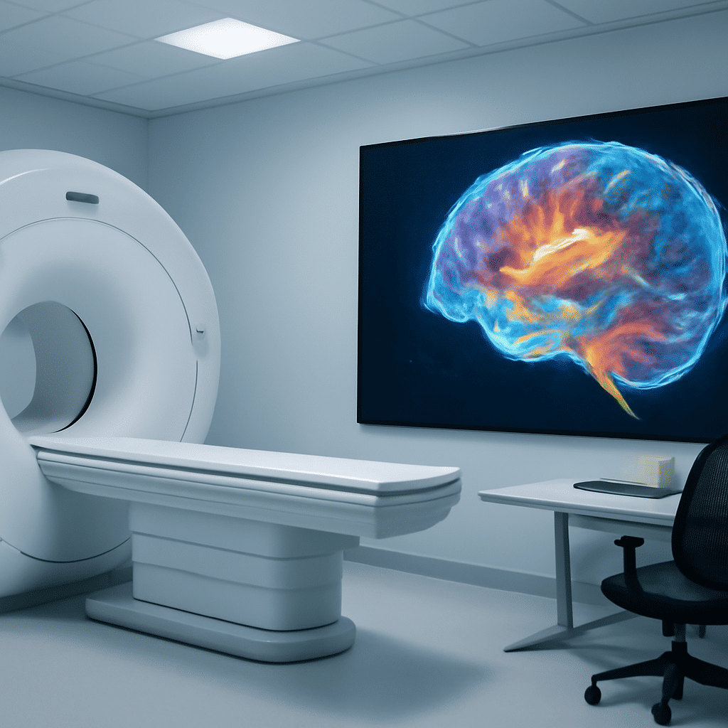
Recent advancements in neuroimaging techniques have revolutionized the way we understand the human brain, offering unprecedented insights into its complex activities and disorders. These state-of-the-art methods have developed to provide non-invasive windows into the brain, allowing researchers and clinicians to observe its workings in remarkable detail.
One of the most significant breakthroughs in neuroimaging is the use of functional MRI (fMRI), which measures brain activity by detecting changes in blood flow. This technique has enabled researchers to map brain activity with high spatial resolution, facilitating a better understanding of how different brain regions coordinate to perform various cognitive tasks. By correlating these patterns with specific mental processes, fMRI has contributed significantly to our knowledge of the brain's functional architecture.
Another cutting-edge technique is diffusion tensor imaging (DTI), which allows scientists to visualize the brain's white matter tracts. This method tracks the diffusion of water molecules along axons, providing insights into the structural connectivity of the brain. By examining these pathways, researchers can better understand how information is transmitted across different brain regions and how alterations in these pathways may be linked to neurological disorders.
Positron emission tomography (PET) remains a powerful tool in neuroimaging as well, enabling the visualization of molecular processes within the brain. By using radiotracers, PET scans can detect various substances such as neurotransmitters or proteins implicated in neurological diseases. This ability to observe biological processes at the molecular level provides crucial information for diagnosis and the development of targeted therapies for conditions like Alzheimer's disease and Parkinson's disease.
Moreover, advancements in magnetoencephalography (MEG) and electroencephalography (EEG) have improved our understanding of the brain's electrical activity. These techniques offer excellent temporal resolution, allowing researchers to capture rapid changes in brain activity in real time. By combining these methods with fMRI and DTI, neuroscientists can gain a more comprehensive picture of brain function and its disruptions in various disorders.
Importantly, these non-invasive techniques have also enabled longitudinal studies, where brain activity and structure can be monitored over time. This is particularly valuable in studying developmental processes and aging, providing critical insights into how the brain changes throughout life and how these changes relate to cognitive functions and disorders.
In clinical settings, advanced neuroimaging techniques are proving invaluable for diagnosing and managing neurological conditions. They provide critical data that can guide surgical interventions, track disease progression, and evaluate treatment efficacy. As researchers continue to refine these technologies and develop new analytical methods, the potential for neuroimaging to transform our understanding of the brain and improve patient outcomes is immense.
In summary, the advancements in neuroimaging are paving the way for a deeper understanding of the brain and its disorders. By offering non-invasive, detailed views of brain activity and structure, these technologies have opened up new avenues for research and clinical application, promising to reshape neuroscience and healthcare in the coming years.